The Layered Approach to Mast Cell Activation Syndrome: Integrating Dr. Afrin’s Framework with Functional Medicine Therapeutics
A Clinical Chapter on Trigger Avoidance, Anaphylaxis Preparedness, Antihistamines, Stabilizers, Antileukotrienes, Omalizumab, LDN, Quercetin, Luteolin, and Methylene Blue in a Stepwise Model
Yoon Hang Kim, MD, MPH
Board-Certified in Preventive Medicine | Integrative & Functional Medicine Physician
Abstract
Mast Cell Activation Syndrome (MCAS) is a multi-system disorder of inappropriate mast cell mediator release whose heterogeneity resists any single-agent solution. This chapter presents a layered, stepwise clinical model that anchors management in the conventional standard of care—trigger identification and avoidance, acute anaphylaxis preparedness, H1/H2 antihistamines, mast cell stabilizers, antileukotrienes, aspirin, and omalizumab—and then extends it with integrative adjuncts used in functional medicine practice: low-dose naltrexone (LDN), the natural flavonoids quercetin and luteolin, and low-dose methylene blue. Each layer is examined for mechanism, dosing, evidence quality, and its place in a graded escalation, with explicit attention to where biological plausibility outruns clinical data. The model is framed against the diagnostic tension between the narrow Vienna consensus criteria and the broader Afrin–Molderings clinical construct, and situates clonal mast cell disease as a distinct entity warranting hematologic referral.
Keywords: mast cell activation syndrome; layered treatment; trigger avoidance; anaphylaxis; antihistamines; cromolyn; ketotifen; omalizumab; low-dose naltrexone; quercetin; luteolin; methylene blue; diamine oxidase; systemic mastocytosis; integrative medicine
Medical Disclaimer
This chapter is for educational purposes only and does not constitute medical advice, diagnosis, or treatment. Mast Cell Activation Syndrome (MCAS) management must be individualized under the guidance of a qualified healthcare provider. Do not start, stop, or modify any treatment based on this information alone. Always consult your physician before making changes to your care plan.
1. Introduction: Why a Layered Approach Is Necessary
Mast Cell Activation Syndrome (MCAS) is a condition in which mast cells—normally protective immune sentinels stationed throughout connective tissue, mucosal surfaces, and perivascular spaces—become chronically dysregulated, releasing a disproportionate cascade of mediators including histamine, prostaglandins, leukotrienes, tryptase, and cytokines.1,2 The clinical result is a multi-system inflammatory syndrome that can mimic dozens of other conditions, presenting with symptoms spanning the gastrointestinal, dermatologic, cardiovascular, neuropsychiatric, and musculoskeletal systems simultaneously.3
This heterogeneity demands a layered, stepwise treatment strategy—one that builds sequentially, adds one intervention at a time, and allows for careful clinical observation at each stage. This chapter presents such a model, anchoring management in the conventional standard of care and extending it with the functional medicine toolkit I employ at www.directintegrativecare.com—specifically low-dose naltrexone (LDN), the natural flavonoids quercetin and luteolin, and low-dose methylene blue (MB), alongside dietary and root-cause strategies. Two companion overviews—MCAS: A New Epidemic in Plain Sight and A Functional Medicine Approach to MCAS—provide the wider context for the framework developed here.
1.1 A Note on Diagnostic Framing
Before any treatment discussion, a word on diagnostic framing is warranted, because two schools of thought coexist in the mast cell field and this chapter should be read with that distinction in mind. The international consensus group led by Valent and Akin—the “Vienna consensus,” also reflected in the AAAAI Mast Cell Disorders Committee Work Group Report—defines MCAS narrowly, requiring all three of the following: typical episodic symptoms involving at least two organ systems; objective laboratory evidence of mast cell involvement, classically an event-related rise in serum tryptase of at least 20% above the individual’s baseline plus 2 ng/mL (the “20% + 2” rule), or another validated mediator; and a demonstrable response to mast-cell-targeted therapy.1,2 By these criteria, true idiopathic MCAS is uncommon, and a substantial share of clients who suspect MCAS do not formally meet the definition.
A broader clinical construct, associated with Dr. Lawrence Afrin and Dr. Gerhard Molderings, emphasizes the extraordinary heterogeneity of individual mediator profiles and leans more heavily on symptom pattern and therapeutic response when strict tryptase criteria are not met.3,4 This chapter operates primarily within that broader construct, because it reflects the population most often encountered in integrative practice. The distinction matters, however: the layered approach below assumes a client who has been appropriately evaluated and in whom MCAS—or a well-defined mast cell activation phenotype—is a reasonable working diagnosis, not a default label applied to any unexplained multi-system illness. Objective mediator workup should precede, not follow, an escalating medication trial. It is equally important to distinguish non-clonal MCAS from clonal mast cell disease (systemic mastocytosis), which is managed differently and is addressed in Section 9.3.
Dr. Afrin, a hematologist-oncologist widely regarded as a leading clinical authority on mast cell disease, has remarked in interviews that he did not encounter a case bearing reasonable similarity to any previous one until he had seen roughly seventy MCAS clients—an anecdotal observation rather than a published statistic, but one that captures the biological variability at the core of this condition. His guiding principles of patience, persistence, and a methodical approach remain a widely respected standard, extended here by immunomodulatory agents, natural mast cell stabilizers, and mitochondrial support that address proposed root-cause drivers of mast cell dysfunction.
2. Foundational Principle: Trigger Identification and Avoidance
Before any pharmacologic layer, the foundation of MCAS management—named in every consensus review—is the systematic identification and avoidance of activation triggers.2,4 Antihistamines and stabilizers are far less effective in a client who continues daily exposure to their principal triggers. Common triggers include specific foods and high-histamine dietary patterns, alcohol, temperature extremes, physical exertion, emotional stress, fragrances and chemical exposures, certain medications and excipients, and, in susceptible clients, hormonal fluctuations and mold or biotoxin exposure. Because triggers are highly individual, a structured symptom-and-exposure diary is often the single most productive early intervention.
This foundational work intersects with the root-cause evaluation that distinguishes the functional medicine approach—gut dysbiosis, chronic infection, mold and biotoxin illness, and hormonal drivers. Several of these threads are developed in companion reviews: CIRS vs. MCAS, Estrogen and MCAS, the Lactobacillus rhamnosus GG gut review, and The COMT Gene and MCAS.
3. Acute Safety: Anaphylaxis Recognition and Epinephrine
No treatment discussion is complete—or safe—without addressing acute anaphylaxis, the most dangerous manifestation of mast cell activation. Any client with a history of, or meaningful risk for, systemic reactions should be prescribed epinephrine auto-injectors, be taught to use them, and carry two doses at all times. Epinephrine is the first-line pharmacotherapy for anaphylaxis; antihistamines and corticosteroids are adjuncts that must never delay or replace it.5 Every such client should have a written anaphylaxis emergency action plan, and severe reactions warrant emergency evaluation with a period of observation for biphasic recurrence.5
This acute-safety layer sits beneath the entire chronic-management framework. The daily regimen described below aims to reduce the frequency and severity of activation over time, but it does not substitute for rescue epinephrine in an acute event. Clients should also be counseled to avoid, where feasible, medications that can worsen anaphylaxis outcomes—such as non-selective beta-blockers—in consultation with their prescribing physicians.
4. Layer 1: Dual Histamine Receptor Blockade
With triggers addressed and acute safety in place, every published treatment algorithm for MCAS begins pharmacologically with dual histamine receptor blockade—the pharmacologic floor upon which subsequent layers are built.2,4 Histamine acts on four receptor subtypes (H1 through H4), but currently available therapeutics target only H1 and H2. By blocking both receptor classes simultaneously, clinicians achieve broader coverage of histamine-mediated symptoms including flushing, pruritus, urticaria, tachycardia, gastrointestinal distress, and headache.
A point of mechanistic precision is warranted here, because a claim circulating widely in the integrative MCAS literature is stated backwards. It is often asserted that famotidine stabilizes mast cells because H2 receptors sit on the mast cell surface. Human mast cells do express H2 receptors6—but H2 signaling on the mast cell raises intracellular cAMP and inhibits mediator release, functioning as a negative-feedback brake. Blocking that receptor does not stabilize the mast cell. The sound rationale for H2 blockade in MCAS is different and still compelling: H2 receptors are widely distributed on target tissues throughout the gastrointestinal tract, cardiovascular system, and elsewhere, so H2 antagonism blocks a large share of histamine’s downstream effects that H1 blockade leaves untouched.7 A proton pump inhibitor does not substitute, because it suppresses acid production rather than blocking histamine at its receptor.
Second-generation H1 antihistamines such as cetirizine or fexofenadine are preferred over first-generation agents (diphenhydramine, hydroxyzine) due to their superior side effect profiles and longer half-lives.2 MCAS clients frequently require doses at two to four times FDA-approved levels under medical supervision.4 These medications work prophylactically—once histamine-mediated symptoms appear, receptor binding has already occurred, and the therapeutic window has been missed.
This foundational layer alone provides meaningful symptom reduction in many MCAS clients, and Dr. Afrin has observed that a fortunate subset achieves satisfactory control with H1/H2 blockade alone. For the majority, this layer establishes a baseline from which additional agents are methodically introduced. Histamine dysregulation also carries under-recognized neuropsychiatric consequences, explored in the companion review The Neuropsychiatric Impact of Histamine Dysregulation in MCAS.
5. Layer 2: Mast Cell Stabilization — Ketotifen and Cromolyn Sodium
When antihistamines alone do not provide adequate control, the next escalation adds agents that prevent mast cell degranulation itself—shifting the strategy from blocking released mediators to preventing their release.
5.1 Ketotifen: Dual-Action Systemic Stabilizer
Ketotifen occupies a unique niche as both a mast cell stabilizer and an H1 antihistamine. Its stabilizing action involves inhibition of calcium influx across mast cell membranes, reducing release of histamine, tryptase, prostaglandins, and other mediators; its H1 antagonism adds histamine blockade beyond cetirizine or fexofenadine.4 A fuller treatment of the evidence appears in the companion review Ketotifen for MCAS.
In functional medicine practice, ketotifen is frequently used as a bridge therapy—providing symptom control while deeper root causes such as gut dysbiosis, mold exposure, chronic infections, or heavy metal burden are identified and addressed. It is available in the United States primarily through compounding pharmacies, as the oral formulation is not commercially marketed domestically. Common starting doses range from 0.5 to 1 mg at bedtime, with titration guided by clinical response and side effects (primarily initial sedation). Maximum therapeutic effect typically requires six to twelve weeks of consistent use—a timeline that reinforces the emphasis on patience.
5.2 Cromolyn Sodium: The GI-Focused Stabilizer
Cromolyn sodium remains a first-line option for GI-predominant MCAS presentations. Its negligible systemic absorption (less than one percent reaches the circulation) means it acts almost exclusively at the gastrointestinal mucosal surface, where a high concentration of mast cells resides. This makes it particularly effective for abdominal pain, nausea, bloating, diarrhea, and food-related activation, though some clients report broader benefit.2 It requires slow introduction, often beginning at one-quarter of an ampule and titrating over weeks; consistent with consensus guidance, full benefit may take four to eight weeks and it should be trialed for at least a month before judging response.2 It is available as an oral solution (Gastrocrom) or compounded into capsules for clients sensitive to the liquid excipients.
6. The Conventional Escalation Tier: Antileukotrienes, Aspirin, Omalizumab, and Corticosteroids
Before the integrative layers, it is essential to place the conventional escalation agents that published MCAS algorithms reach for after antihistamines and stabilizers.2 Omitting these would misrepresent the standard of care; the integrative layers that follow are adjuncts to this tier, not replacements for it. This tier sits between Layer 2 and Layer 3 in the sequence of escalation.
Leukotriene pathway modulation. When symptoms persist despite H1/H2 blockade and stabilizers—particularly wheezing, nasal congestion, and some gastrointestinal and vascular symptoms—leukotriene receptor antagonists such as montelukast, or the 5-lipoxygenase inhibitor zileuton, target the cysteinyl leukotriene arm of mediator release that antihistamines do not touch.2
Prostaglandin modulation. For flushing and other symptoms driven predominantly by prostaglandin D2, aspirin or other COX inhibitors are used in clients who tolerate them—with caution, because aspirin can itself provoke reactions in a subset of mast cell clients. This is a targeted intervention against a specific mediator pathway the antihistamine foundation leaves unaddressed.2,4
Omalizumab. For refractory disease, particularly presentations dominated by recurrent anaphylaxis, the anti-IgE monoclonal antibody omalizumab reduces mast cell activatability and is the best-supported escalation step in the conventional literature. A client with refractory, anaphylaxis-predominant MCAS belongs on a trial of omalizumab before or alongside any integrative adjunct.2
Corticosteroids. Systemic steroids can be effective for severe or refractory flares but are reserved for short courses because of their considerable adverse-effect burden with chronic use.2
With this conventional tier explicit, the integrative layers below address complementary axes of mast cell biology—immune signaling, natural mediator stabilization, and mitochondrial and vascular support—and are introduced alongside or after these agents, not in their place.
7. Layer 3: Immunomodulation — Low-Dose Naltrexone (LDN)
This layer shifts from mediator blockade and stabilization to upstream immunomodulation. Low-dose naltrexone is a biologically plausible adjunct operating through pathways distinct from any antihistamine or stabilizer.8 It should be stated at the outset that LDN does not appear in the major consensus MCAS algorithms—it is absent from the AAAAI work group report and the Vienna consensus1,2—so its use here is an extrapolation from adjacent immunologic literature and a growing but uncontrolled clinical signal, not a guideline-endorsed step.
At doses of 0.5 to 4.5 mg daily (typically at bedtime), LDN functions as a short-acting mu-opioid antagonist that transiently blocks opioid receptors. This paradoxical blockade is proposed to trigger a compensatory upregulation of endogenous endorphins and enkephalins, which are thought to interact with regulatory T cells, influencing T- and B-lymphocyte activity and recalibrating the adaptive immune response.8
The mechanisms by which LDN may benefit MCAS are inferred from adjacent literature rather than demonstrated in MCAS clients directly. LDN antagonizes Toll-like receptor 4 (TLR4), whose activation drives NF-κB-mediated inflammatory cascades; TLR4 is expressed on mast cells and microglia alike, and its inhibition reduces pro-inflammatory cytokines including IL-6 and TNF-α while promoting anti-inflammatory mediators such as IL-10.8 LDN also modulates microglial activation, relevant for clients with neuroinflammation, brain fog, and neuropsychiatric symptoms.8,9 A further proposed pathway is that T-cell microparticles activate mast cells and that LDN’s reduction of excessive T-cell dysfunction disrupts this loop—this remains a hypothesis, and Weinstock, who proposed it, has been explicit that the existence of opioid receptors on mast cells themselves is not clear.10
The clinical evidence base consists of case reports, small case series, and uncontrolled clinical audits rather than randomized controlled trials, and the figures deserve to be stated precisely. In the frequently cited 2018 BMJ Case Report by Weinstock and colleagues, a client with severe POTS and MCAS received ultra-low-dose naltrexone at 1 mg nightly; after six weeks this produced a 7% decrease in POTS severity scores and a 17% decrease in MCAS severity scores, with improvement in body pain, mood, memory, sleep, flushing, odor and food sensitivities, and paresthesia.10 Larger gains followed the subsequent addition of IVIg and rifaximin, so the composite response cannot be attributed to naltrexone alone.
A figure widely quoted as “LDN Research Trust data” is more accurately Dr. Leonard Weinstock’s own unpublished clinical audit, presented in a video interview hosted by the Trust. In that series of 116 MCAS clients, 60% reported some improvement, 20% reported no effect, and 20% discontinued because of side effects—unsurprising given how frequently MCAS clients react to medications and excipients. Within the 70 clients who improved, the symptom-level numbers are small: abdominal pain improved in 15, joint pain and fatigue in 11 each, muscle pain in 9, diarrhea in 6, constipation and restless legs in 5 each, and reduction in mast cell flares specifically in only 4. Because this audit is unpublished, it should be cited as such and weighted accordingly.
A 2025 clinical audit from University Hospitals of Leicester provides a second uncontrolled data point: 40 MCAS clients treated with LDN showed improvement in joint pain, fatigue, and gastrointestinal and skin symptoms, while 20 did not tolerate the medication.11 This report appears as a mini-review in a low-circulation journal and has not been independently replicated, so it should be weighted as supportive rather than confirmatory. All were already receiving antihistamines and mast cell stabilizers before LDN was added—consistent with the layered model here, in which LDN is an addition to a foundation, not a replacement for it.
In my practice, LDN is positioned at Layer 3 because it addresses a different axis of MCAS pathophysiology—immune dysregulation—rather than simply blocking mediators or stabilizing membranes, and it is offered as a complement to, not a substitute for, the conventional escalation agents above. For a detailed mechanistic review, see Why Low-Dose Naltrexone Calms Mast Cells.
8. Layer 4: Natural Mast Cell Stabilizers — Quercetin and Luteolin
This layer incorporates natural flavonoids providing mast cell stabilization through mechanisms complementary to pharmaceutical agents. Quercetin and luteolin are the two most extensively studied natural mast cell stabilizers and are widely used in integrative MCAS management.
8.1 Quercetin
Quercetin, a bioflavonoid found in onions, apples, berries, and green tea, inhibits release of histamine, tryptase, and pro-inflammatory cytokines from mast cells via modulation of intracellular calcium signaling, inhibition of NF-κB activation, and suppression of protein kinase C pathways involved in degranulation.12 It also inhibits prostaglandin and leukotriene release, addressing mediator pathways beyond histamine.12,13
The primary limitation is poor oral bioavailability—absorption of standard formulations is low enough that human plasma concentrations fall well below those used in the cell-culture experiments that demonstrate mast cell inhibition. This gap between in vitro potency and in vivo exposure is the central caveat for all flavonoid therapy.13 Liposomal or phytosome-complexed preparations improve absorption and are preferred. Typical dosing ranges from 500 to 1,000 mg daily, sometimes combined with vitamin C for general antioxidant support. The favorable safety profile makes quercetin a rational addition, particularly for clients sensitive to pharmaceutical agents.
8.2 Luteolin
Luteolin, found in celery, chamomile, green peppers, and parsley, shares quercetin’s stabilizing properties but offers advantages for neuroinflammatory presentations. It inhibits mast cell activation by suppressing intracellular calcium influx and blocking protein kinase C activation—the same signaling pathways through which the related flavonols act.12 It is worth noting that the published head-to-head comparison against cromolyn was conducted with quercetin, not luteolin13; claims that luteolin specifically outperforms cromolyn are frequently repeated but are extrapolated rather than directly demonstrated.
What distinguishes luteolin is its reported ability to cross the blood-brain barrier, making it of particular interest for clients with neuroinflammation, brain fog, and cognitive dysfunction, as it may modulate CNS mast cells and reduce microglial activation—a body of work advanced substantially by Theoharides and colleagues.9,14 In practice, quercetin and luteolin are often combined for complementary systemic and CNS coverage; liposomal formulations are preferred, and typical luteolin dosing ranges from 100 to 300 mg daily. For a comprehensive review, see Luteolin for MCAS: A Natural Mast Cell Stabilizer Worth Understanding.
9. Layer 5: Mitochondrial and Neuroimmune Support — Methylene Blue
The final layer introduces methylene blue (MB), which occupies the most speculative position in this framework. It is included because it addresses a mitochondrial dimension of mast cell dysfunction that no conventional antihistamine, stabilizer, or immunomodulator directly targets—but its supporting evidence in MCAS is the thinnest of any agent here, and one of its proposed mechanisms may not act in the direction commonly assumed.
Mast cells depend on mitochondrial function for both mediator synthesis and degranulation signaling. When mitochondrial function is compromised—by oxidative stress, chronic infection, environmental toxin exposure, or genetic polymorphisms—mast cells may become more reactive and less responsive to stabilizing interventions. This is the proposed rationale for mitochondrial support, though it remains a hypothesis rather than an established MCAS mechanism.
At the low oral doses used in integrative practice—generally 5 to 20 mg daily, or roughly 0.1 to 0.3 mg/kg, well below the milligram-per-kilogram dosing used acutely for methemoglobinemia—methylene blue acts as an alternative electron carrier in the mitochondrial electron transport chain, shuttling electrons between Complex I and Complex III, supporting ATP production while reducing reactive oxygen species.15 This dual action has been proposed to support cellular resilience, though the supporting studies were conducted in non-mast-cell systems.15
Beyond its mitochondrial effects, methylene blue inhibits soluble guanylate cyclase and thereby the nitric oxide–cyclic GMP (NO–cGMP) pathway. This is the mechanism behind its established acute use—as a rescue agent for refractory vasoplegic and anaphylactic hypotension, where blocking NO-driven vasodilation restores vascular tone. In the POTS–MCAS overlap, the same mechanism may blunt the downstream vascular consequences of mediator release: flushing, vasodilation, and vasomotor instability.
A crucial mechanistic caveat must be stated plainly, however. Reducing these vascular effects is not the same as stabilizing the mast cell, and the direction of the NO–cGMP pathway’s effect on mast cells themselves is not settled. At least one in vivo microcirculatory study found that guanylate cyclase inhibition—by methylene blue specifically—increased perivascular mast cell degranulation rather than suppressing it, mirroring the effect of nitric oxide synthase blockade.16 In other words, the NO–cGMP axis may restrain degranulation in some vascular beds, so blocking it could, in principle, promote mediator release even as it reduces the vascular response to that release. The honest position is that methylene blue may help vasomotor symptoms through a hemodynamic mechanism while its effect on the mast cell itself remains uncertain and possibly unfavorable.
Methylene blue has also demonstrated neuroprotective properties in preclinical models—crossing the blood-brain barrier and reducing microglial-mediated neuroinflammation—making it of theoretical interest alongside luteolin. Taken together, the absence of direct MCAS evidence, a mitochondrial rationale drawn largely from non-mast-cell models, and an unsettled (possibly unfavorable) effect on the mast cell place methylene blue at the most experimental end of this framework. It should be reserved, if used at all, for carefully selected clients after conventional and better-supported options have been exhausted. For a full review, see Low Dose Methylene Blue in Functional Medicine.
Methylene blue also carries important precautions. It is contraindicated with serotonergic medications (SSRIs, SNRIs, MAOIs) due to the risk of serotonin syndrome. G6PD deficiency must be ruled out prior to initiation. Clients should be informed that MB will turn urine blue-green—a harmless but sometimes alarming effect.
10. Adjunctive and Root-Cause Supportive Therapies
Beyond the graded pharmacologic and integrative layers, several supportive strategies are routinely woven into functional medicine MCAS care. These are best understood as adjuncts that lower total mediator burden or address upstream drivers, not as stand-alone treatments.
10.1 Dietary Modification and Diamine Oxidase (DAO)
A low-histamine diet is a mainstay for clients whose symptoms track with food, reducing the enteral histamine load that a compromised gut may fail to metabolize.17 It should be implemented thoughtfully and time-limited: overly restrictive elimination risks nutritional inadequacy and disordered eating, and consensus guidance recommends involving a dietitian experienced in food intolerance. Diamine oxidase (DAO) is the principal enzyme degrading ingested histamine at the intestinal mucosa; exogenous DAO supplementation taken before meals has been proposed as an adjunct in histamine intolerance, and cofactors for DAO activity include vitamin B6, copper, and vitamin C.17 The evidence base for DAO supplementation remains limited and heterogeneous, so it is offered as a reasonable, low-risk trial rather than an established therapy.
10.2 Vitamin D Repletion
Vitamin D acts as an immunomodulatory hormone with effects on mast cell biology, and deficiency is common in chronically ill MCAS clients. Correcting a documented deficiency is a sensible, low-risk supportive measure. The mechanistic case is developed in the companion review Vitamin D as a Mast Cell–Modulating Hormone.
10.3 Gut Microbiome and Hormonal Modifiers
Because the gut is both a major reservoir of mast cells and a site of histamine metabolism, targeted microbiome support may reduce activation; the strain-specific evidence is reviewed in Lactobacillus rhamnosus GG (LGG) in MCAS. Hormonal fluctuations, particularly estrogen, can modulate mast cell reactivity and account for catamenial symptom patterns—explored in Estrogen and MCAS—while genetic modifiers such as COMT may influence individual presentation, as discussed in The COMT Gene and MCAS.
11. Special Clinical Situations
11.1 Procedural Premedication
Clients with significant MCAS are at elevated risk of activation during surgery, endoscopy, or radiocontrast administration. Premedication protocols—typically H1 and H2 antihistamines with a corticosteroid, and in selected clients a benzodiazepine—are used before such procedures to reduce the risk of perioperative reactions, in coordination with the anesthesia and procedural teams.2,4
11.2 Benzodiazepines
Benzodiazepines have been proposed as an adjunct in mast cell activation disease, on the rationale that mast cells express benzodiazepine binding sites and that anxiety frequently accompanies—and can itself trigger—activation.4 The direct evidence for a mast-cell-stabilizing effect is limited, and given dependence potential these agents are best used judiciously and situationally, principally for procedural anxiety, rather than as a maintenance layer.
11.3 Clonal Mast Cell Disease and Cytoreduction
When evaluation confirms clonal disease—systemic mastocytosis, typically driven by the KIT D816V mutation—management diverges from the non-clonal model that is the focus of this chapter, and hematologic referral is warranted.1 Options in this setting extend to cytoreductive therapy with hydroxyurea (hydroxycarbamide)18 and, for advanced disease, KIT-targeted tyrosine kinase inhibitors such as midostaurin and avapritinib.19 Small case series have also explored low-dose naltrexone alongside hydroxycarbamide in mast cell disorders11, but the cytoreductive and KIT-targeted agents are hematology-directed therapies beyond the scope of an integrative model and are noted here for completeness and appropriate referral.
12. Clinical Algorithm: Matching Layers to Client Phenotype
Figure 1 summarizes the complete model. Trigger avoidance forms the base, the pharmacologic layers ascend from it in order of escalation, and acute-safety preparedness runs parallel to every layer rather than sitting within the sequence. Evidence strength decreases with height.
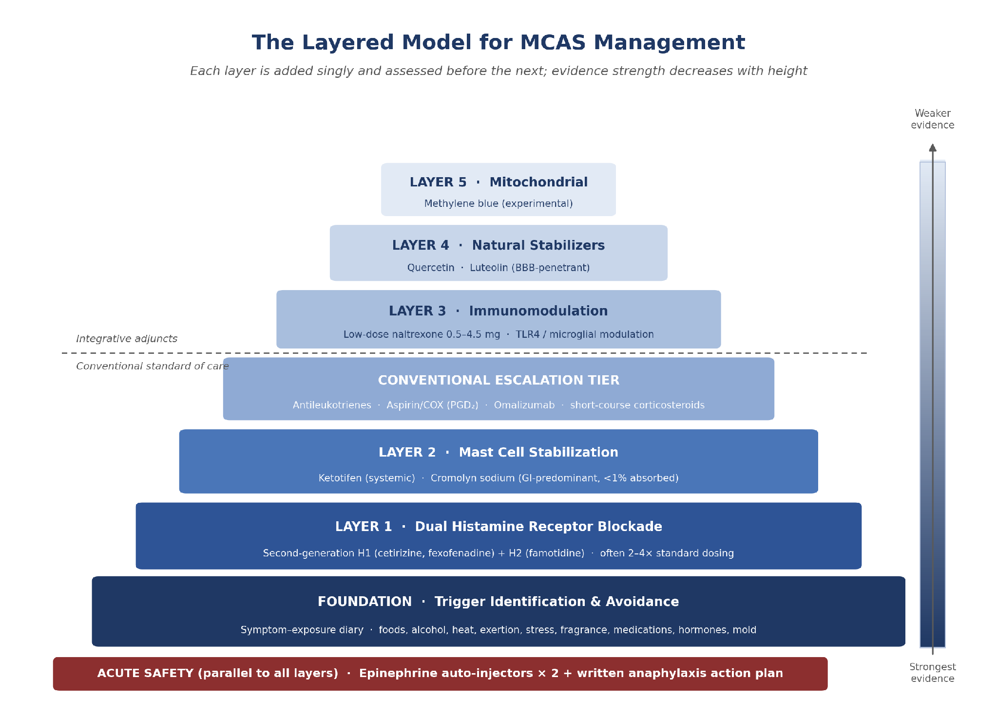
Figure 1. The layered model for MCAS management. The dashed divider separates guideline-endorsed conventional therapy from integrative adjuncts whose evidence base is largely mechanistic or uncontrolled.
The model is not rigidly sequential for every client. The clinical art lies in identifying the predominant phenotype and selecting the most relevant layers, with the conventional escalation tier available as symptoms warrant. Two elements apply to every phenotype below: (1) trigger identification and avoidance, and (2) an epinephrine auto-injector and written action plan for any client at risk of anaphylaxis.
Table 1. Phenotype-guided application of the model shown in Figure 1. Adapted from clinical algorithms published at ifmsynergy.com/tag/mcas and aligned with AAAAI work-group escalation steps.² All interventions require individualization under clinical supervision.
13. Guiding Principles: The Afrin Framework Applied
Dr. Afrin’s three principles—patience, persistence, and a methodical approach—are clinical necessities. MCAS does not yield to aggressive polypharmacy or rapid-fire changes. Each layer should be introduced as a single variable, maintained for an adequate trial period (typically four to twelve weeks depending on the agent), and assessed for both benefit and adverse effects before the next is added.
This methodical escalation lets clinicians identify which agents produce benefit or cause reactions, avoids the confounding of simultaneous multi-agent introduction, and respects the exquisite sensitivity many MCAS clients have to new substances—including the medications intended to help them. Even beneficial agents like cromolyn and ketotifen can initially worsen symptoms in hypersensitive individuals if introduced too rapidly.
The model also acknowledges that management is not about perfection. The realistic goal, as Dr. Afrin has articulated, is to help clients feel significantly better than their pre-treatment baseline the majority of the time. Setting that expectation benefits clinicians and clients alike: complete resolution is rarely achievable, but substantial, meaningful improvement is well within reach for most clients who pursue methodical, layered treatment on a foundation of trigger avoidance and acute-safety preparedness.
14. Conclusion
MCAS requires an approach as individualized as the condition itself. The model presented here—trigger avoidance and anaphylaxis preparedness as the foundation, then an antihistamine floor, mast cell stabilization, the conventional escalation tier of antileukotrienes, aspirin, and omalizumab, and then immunomodulation with LDN, natural flavonoid support with quercetin and luteolin, and mitochondrial support with methylene blue—synthesizes the evidence-based pharmacologic framework with the root-cause-oriented perspective of functional medicine, supported by dietary, microbiome, hormonal, and repletion strategies.
No single agent addresses all dimensions of mast cell dysregulation. Histamine blockade alone does not prevent degranulation. Stabilizers alone do not modulate upstream immune signaling. Immunomodulators alone do not address mitochondrial and vascular contributions. It is the layered integration of these mechanisms—anchored in the conventional standard of care and extended thoughtfully by integrative adjuncts—that offers clients the most comprehensive path toward stabilization and improved quality of life.
The evidence base is uneven, and honesty about that gradient is part of good care. The trigger-avoidance, epinephrine, antihistamine, stabilizer, antileukotriene, and omalizumab layers rest on the firmest ground; LDN carries a plausible mechanism and early uncontrolled signal; the flavonoids are supported mainly by preclinical data; and methylene blue remains frankly experimental for this indication. Clinicians and clients must hold the tension between biological plausibility and the current limits of rigorous trial data. What is clear is that MCAS is treatable, that most clients can achieve meaningful improvement, and that a methodical, layered approach built on the conventional foundation remains the most reliable path to that outcome.
Related Reading: The IFM Synergy MCAS Series
The following companion reviews expand on specific threads referenced throughout this chapter:
• MCAS: A New Epidemic in Plain Sight
• A Functional Medicine Approach to MCAS
• Why Low-Dose Naltrexone Calms Mast Cells
• Luteolin for MCAS: A Natural Mast Cell Stabilizer Worth Understanding
• Low Dose Methylene Blue in Functional Medicine
• Vitamin D as a Mast Cell–Modulating Hormone
• Lactobacillus rhamnosus GG (LGG) in MCAS
• The Neuropsychiatric Impact of Histamine Dysregulation in MCAS
References
1. Valent P, Akin C, Arock M, Brockow K, Butterfield JH, Carter MC, et al. Definitions, criteria and global classification of mast cell disorders with special reference to mast cell activation syndromes: a consensus proposal. Int Arch Allergy Immunol. 2012;157(3):215–225. doi:10.1159/000328760
2. Weiler CR, Austen KF, Akin C, Barkoff MS, Bernstein JA, Bonadonna P, et al. AAAAI Mast Cell Disorders Committee Work Group Report: Mast cell activation syndrome (MCAS) diagnosis and management. J Allergy Clin Immunol. 2019;144(4):883–896. doi:10.1016/j.jaci.2019.08.023
3. Afrin LB, Butterfield JH, Raithel M, Molderings GJ. Often seen, rarely recognized: mast cell activation disease—a guide to diagnosis and therapeutic options. Ann Med. 2016;48(3):190–201. doi:10.3109/07853890.2016.1161231
4. Molderings GJ, Haenisch B, Brettner S, Homann J, Menzen M, Dumoulin FL, Panse J, Butterfield J, Afrin LB. Pharmacological treatment options for mast cell activation disease. Naunyn-Schmiedeberg’s Arch Pharmacol. 2016;389(7):671–694. doi:10.1007/s00210-016-1247-1
5. Shaker MS, Wallace DV, Golden DBK, Oppenheimer J, Bernstein JA, Campbell RL, et al. Anaphylaxis—a 2020 practice parameter update, systematic review, and Grading of Recommendations, Assessment, Development and Evaluation (GRADE) analysis. J Allergy Clin Immunol. 2020;145(4):1082–1123. doi:10.1016/j.jaci.2020.01.017
6. Lippert U, Artuc M, Grützkau A, Babina M, Guhl S, Haase I, et al. Human skin mast cells express H2 and H4, but not H3 receptors. J Invest Dermatol. 2004;123(1):116–123. doi:10.1111/j.0022-202X.2004.22721.x
7. Cardet JC, Castells MC, Hamilton MJ. Immunology and clinical manifestations of non-clonal mast cell activation syndrome. Curr Allergy Asthma Rep. 2013;13(1):10–18. doi:10.1007/s11882-012-0326-8
8. Toljan K, Vrooman B. Low-dose naltrexone (LDN)—review of therapeutic utilization. Med Sci (Basel). 2018;6(4):82. doi:10.3390/medsci6040082
9. Theoharides TC, Tsilioni I, Bawazeer M. Mast cells, neuroinflammation and pain in fibromyalgia syndrome. Front Cell Neurosci. 2019;13:353. doi:10.3389/fncel.2019.00353
10. Weinstock LB, Brook JB, Myers TL, Goodman B. Successful treatment of postural orthostatic tachycardia and mast cell activation syndromes using naltrexone, immunoglobulin and antibiotic treatment. BMJ Case Rep. 2018;2018:bcr-2017-221405. doi:10.1136/bcr-2017-221405
11. Myers B, et al. Use of low dose naltrexone and hydroxycarbamide for mast cell disorders (ISM, MCAS, HaT). J Cancer Prev Curr Res. 2025;16(5):134–135. [Mini review / clinical audit, University Hospitals of Leicester, UK.]
12. Kempuraj D, Madhappan B, Christodoulou S, Boucher W, Cao J, Papadopoulou N, Cetrulo CL, Theoharides TC. Flavonols inhibit proinflammatory mediator release, intracellular calcium ion levels and protein kinase C theta phosphorylation in human mast cells. Br J Pharmacol. 2005;145(7):934–944. doi:10.1038/sj.bjp.0706246
13. Weng Z, Zhang B, Asadi S, Sismanopoulos N, Butcher A, Fu X, Katsarou-Katsari A, Antoniou C, Theoharides TC. Quercetin is more effective than cromolyn in blocking human mast cell cytokine release and inhibits contact dermatitis and photosensitivity in humans. PLoS One. 2012;7(3):e33805. doi:10.1371/journal.pone.0033805
14. Theoharides TC. Luteolin as a therapeutic option for multiple sclerosis. J Neuroinflammation. 2009;6:29. doi:10.1186/1742-2094-6-29
15. Xiong ZM, Choi JY, Wang K, et al. Methylene blue alleviates nuclear and mitochondrial abnormalities in progeria. Aging Cell. 2016;15(2):279–290. doi:10.1111/acel.12434 [Cited for methylene blue’s mitochondrial-antioxidant mechanism; this study examined progeria fibroblasts, not mast cells.]
16. Kimura M, Mitani H, Bandoh T, Totsuka T, Hayashi S. Mast cell degranulation in rat mesenteric venule: effects of L-NAME, methylene blue and ketotifen. Pharmacol Res. 1999;39(5):397–402. doi:10.1006/phrs.1999.0451 [In vivo microcirculatory study in which guanylate cyclase inhibition by methylene blue increased perivascular mast cell degranulation.]
17. Comas-Basté O, Sánchez-Pérez S, Veciana-Nogués MT, Latorre-Moratalla M, Vidal-Carou MDC. Histamine intolerance: the current state of the art. Biomolecules. 2020;10(8):1181. doi:10.3390/biom10081181
18. Weinstock LB, Brook JB, Molderings GJ. Efficacy and toxicity of hydroxyurea in mast cell activation syndrome patients refractory to standard medical therapy: retrospective case series. Naunyn-Schmiedeberg’s Arch Pharmacol. 2022;395(11):1441–1447. doi:10.1007/s00210-022-02282-8
19. Gotlib J, Kluin-Nelemans HC, George TI, Akin C, Sotlar K, Hermine O, et al. Efficacy and safety of midostaurin in advanced systemic mastocytosis. N Engl J Med. 2016;374(26):2530–2541. doi:10.1056/NEJMoa1513098
Note on evidence quality: References were individually verified against primary sources. The trigger-avoidance, epinephrine, antihistamine, stabilizer, antileukotriene, and omalizumab layers are the best supported. LDN evidence in MCAS consists of case reports, case series, and registry data rather than randomized trials; the 116-client Weinstock series cited in Section 7 is an unpublished clinical audit and is identified as such in the text. Flavonoid data (quercetin, luteolin) derive predominantly from in vitro and preclinical models. The methylene blue mitochondrial literature is largely non-mast-cell, and at least one in vivo study suggests its guanylate cyclase inhibition may promote rather than suppress perivascular mast cell degranulation. Cytoreductive and KIT-targeted agents (hydroxyurea, midostaurin, avapritinib) apply to clonal mast cell disease, not non-clonal MCAS. No integrative agent discussed here is FDA-approved for MCAS.
About Dr. Kim
Dr. Yoon Hang “John” Kim is a board-certified physician with over 20 years of clinical experience. A graduate of the University of Arizona Integrative Medicine Fellowship (Osher Fellow under Dr. Andrew Weil), he holds board certifications in Preventive Medicine and Integrative & Holistic Medicine, along with UCLA medical acupuncture certification and IFM Scholar status. He specializes in low-dose naltrexone (LDN), autoimmune conditions, chronic pain, integrative oncology, fibromyalgia, chronic fatigue syndrome, mast cell activation syndrome, and mold toxicity. He is the author of 3 books and over 20 peer-reviewed articles.
Professional: www.yoonhangkim.com | Clinical: www.directintegrativecare.com


