MCAS as a Common Node of Inflammatory Hyperreactivity:
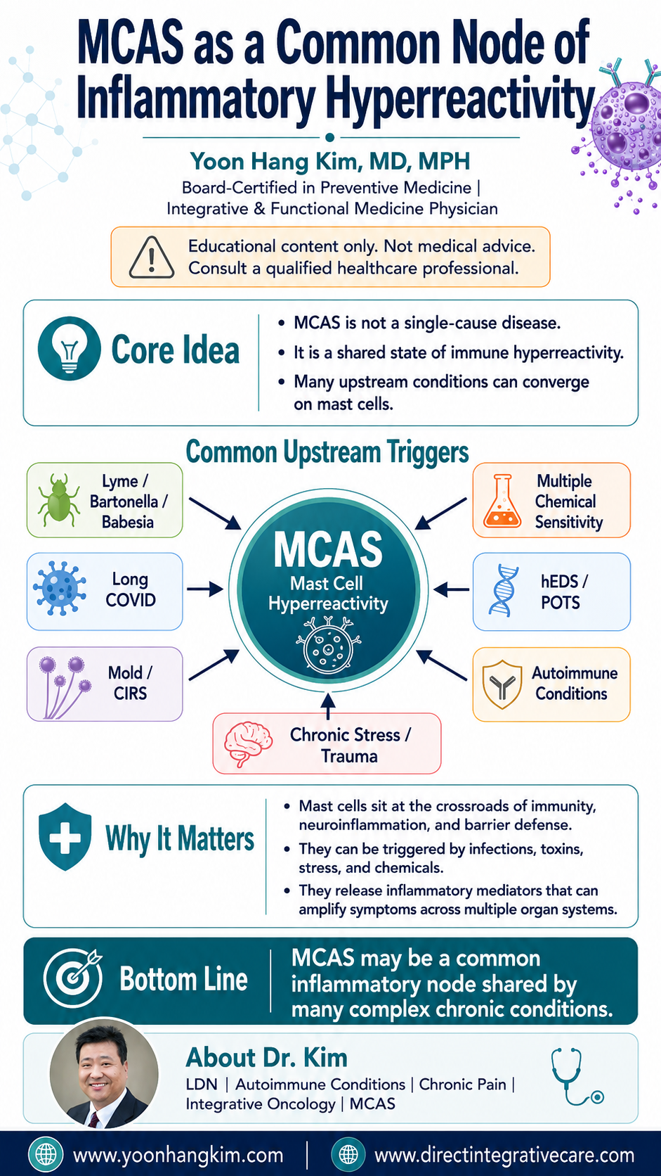
How Multiple Upstream Triggers Converge on Mast Cell Dysregulation
Yoon Hang Kim, MD, MPH
Board-Certified in Preventive Medicine | Integrative & Functional Medicine Physician
Disclaimer: This content is for educational and informational purposes only and does not constitute medical advice, diagnosis, or treatment. Always consult a qualified healthcare provider for individualized medical guidance.
Introduction
In the landscape of complex chronic illness, a clinical pattern has become increasingly difficult to ignore: clients with profoundly different index diagnoses—Lyme disease, Long COVID, mold illness, chemical sensitivity, connective tissue disorders—present with remarkably similar multi-system, fluctuating, histamine-driven symptom profiles. The conventional approach of siloing these conditions into separate specialties has left many clients under-recognized and under-treated. A more useful framework is emerging, one rooted in immunology and clinical pattern recognition: Mast Cell Activation Syndrome (MCAS) as a shared downstream state—a common inflammatory node—that multiple upstream triggers converge upon.
This is not a claim that every complex chronic illness is MCAS. Rather, it is the recognition that mast cells, by virtue of their unique immunological position and activation diversity, are particularly vulnerable to becoming chronically dysregulated by a wide range of upstream insults—and that once dysregulated, they function as inflammatory amplifiers that generate a clinically recognizable phenotype regardless of the original trigger.
Why Mast Cells Occupy This Unique Position
Mast cells are tissue-resident immune cells found at virtually every interface between the body and its environment: the skin, gut mucosa, respiratory epithelium, meninges, and perivascular spaces throughout every organ system. They function as immunological sentinels, positioned to detect and respond to both external threats and internal distress signals. As Akin (2017) described in a comprehensive review, mast cells are “designed to detect and respond to triggers of internal or external stress or danger,” and some degree of mast cell activation is physiologic and necessary for homeostasis [1].
What distinguishes mast cells from other immune cells—and what makes them uniquely suited to serve as a convergence point—is their extraordinary activation diversity. Unlike lymphocytes, which require antigen-specific priming, mast cells can be activated through multiple non-IgE pathways: pathogen-associated molecular patterns (PAMPs), cytokines, complement components, neuropeptides (including corticotropin-releasing hormone and substance P), physical stimuli, temperature changes, hormonal fluctuations, and psychological stress [1, 2]. Petra, Theoharides, and colleagues (2014) catalogued this broad activation profile, noting that mast cells respond to “cytokines, environmental, food, infectious, drug and stress triggers,” releasing multiple mediators that produce multi-system symptoms [2].
Upon activation, mast cells release a remarkably broad repertoire of mediators—including histamine, tryptase, prostaglandins, leukotrienes, heparin, cytokines (TNF-α, IL-6, IL-1β), and vascular endothelial growth factor—that amplify inflammation across every organ system. The commonly cited figure of “over 200 mediators” is now an underestimate: a systematic compilation by Molderings and Afrin (2023) identified 390 substances that human mast cells can secrete upon activation, and the authors note even this may undercount the true repertoire [3]. Critically, mast cells do not merely respond to inflammation; they perpetuate and amplify it. This amplification effect is central to the common node concept: once mast cells become hyper-reactive, they can generate a self-sustaining inflammatory state that may persist after the original trigger has been addressed.
Upstream Triggers That Converge on the MCAS Node
Tick-Borne Infections
The relationship between Borrelia burgdorferi and mast cell activation was established over two decades ago. Talkington and Nickell (1999) demonstrated that B. burgdorferi spirochetes induce low but detectable mast cell degranulation and stimulate the synthesis and secretion of TNF-α by mast cells in vitro [4]. This was among the first evidence that an infectious pathogen could engage the mast cell activation cascade through non-IgE pathways. Later work by Bernard and colleagues (2017) refined this picture: using primary mouse mast cells, they found that the lipidated outer surface protein OspC induces degranulation, while whole spirochetes produce a different inflammatory profile, and that mast cells did not appear to control bacterial proliferation during primary infection in vivo [5]. The mechanistic relationship is therefore real but more nuanced than a simple direct-activation model. Clinically, the relevant question is whether persistent tick-borne infection can maintain chronic mast cell stimulation — a plausible and frequently observed pattern in practice, though one that remains to be established by controlled human studies.
Long COVID and Post-Acute SARS-CoV-2
The COVID-19 pandemic provided compelling real-time evidence for the MCAS convergence model. Theoharides and Kempuraj (2023) reviewed evidence that the SARS-CoV-2 spike protein can activate brain mast cells and microglia, contributing to neuroinflammation and blood-brain barrier dysfunction in Long COVID [6]. Zhang et al. (2025) elucidated the intracellular signaling mechanism, demonstrating that coronavirus spike proteins activate mast cell degranulation via the Src/PI3K/AKT/Ca²⁺ signaling cascade [7].
The clinical overlap between MCAS and Long COVID was directly quantified by Weinstock et al. (2021), who found that mast cell activation symptoms were significantly increased in Long COVID subjects compared to pre-COVID baselines and general population controls. Remarkably, the symptom profile and severity in Long COVID subjects closely mimicked that of established MCAS clients [8]. The authors also noted that low dose naltrexone (LDN) has been used in both MCAS and Long COVID, proposing that its benefit may come from dampening T-cell cytokine signaling that activates mast cells and from blocking Toll-like receptors on mast cells and microglia [8].
Mold Illness and CIRS
Mycotoxins and mold-associated inflammagens activate the innate immune system, and mast cells are among the cell populations that respond to these exposures. The Shoemaker model of chronic inflammatory response syndrome (CIRS) describes a biotoxin-driven illness in HLA-susceptible individuals whose impaired clearance mechanisms are proposed to lead to chronic immune activation. This section warrants an explicit caveat that the others do not: unlike the tick-borne, viral, chemical, and neuroendocrine pathways discussed above, the CIRS framework has not been validated in the peer-reviewed immunology literature to a comparable standard, and the HLA-susceptibility and biotoxin-clearance components remain contested. What can be said with more confidence is descriptive: mold-exposed clients frequently present with symptom patterns consistent with mast cell activation, and in clinical practice mast cell stabilization is often a useful component of their care alongside exposure remediation. The mechanistic account should be held more loosely than the clinical observation.
Multiple Chemical Sensitivity and the TILT Model
The relationship between chemical intolerance and mast cell activation has been formalized through the work of Miller et al. (2021), who proposed that toxicant-induced loss of tolerance (TILT) provides a mechanism for chemical intolerance (CI) that operates through mast cell sensitization [9]. TILT describes a two-stage disease process: initiation, in which a major exposure or series of lower-level exposures sensitizes mast cells, and triggering, in which previously tolerated chemicals, foods, and medications provoke multi-system symptoms through mast cell degranulation. Using the validated Quick Environmental Exposure and Sensitivity Inventory (QEESI), the investigators compared 147 clients diagnosed with MCAS against 345 individuals reporting chemical intolerance and 76 healthy controls, finding close correspondence between the symptom patterns of the MCAS and TILT groups [9]. The authors concluded that xenobiotic-driven mast cell activation is a plausible unifying biological mechanism for CI/TILT — a hypothesis-generating finding rather than a demonstration of causation.
Hypermobile Ehlers-Danlos Syndrome (hEDS)
The co-occurrence of hEDS, MCAS, and postural orthostatic tachycardia syndrome (POTS)—the so-called “trifecta” or “triad”—has become one of the most recognized clinical phenotypes in complex chronic illness. Seneviratne, Maitland, and Afrin (2017) reviewed mast cell disorders in Ehlers-Danlos syndrome, noting that mast cells reside within the extracellular matrix and that altered connective tissue structure in EDS may change mast cell behavior [10]. The most frequently cited prevalence figure — that 66% of affected individuals show mast cell involvement — warrants careful reading: it derives from a small series (N = 15) of clients who had both hEDS and POTS, and it describes symptoms consistent with mast cell activation rather than formal MCAS diagnostic criteria. It should not be read as “66% of hEDS clients have MCAS.” Separately, whole-genome sequencing work by Shirvani et al. (2024) in a small cohort (18 hEDS participants, 7 first-degree relative controls) identified variants in HLA-B, HLA-DRB1, MT-CYB, HTT, and MUC3A, and reported that 72.2% of hEDS participants had documented mast cell hypersensitivity versus 14.2% of controls [11]. The authors themselves flag the small sample as a significant limitation.
This association is genuinely contested, and intellectual honesty requires saying so plainly. Kohn and Chang (2020) concluded that evidence supporting a unified relationship among the three conditions is largely based on biased or outdated criteria, and that no evidence-based common pathophysiological mechanism has been described [12]. More pointedly, a multicenter retrospective review published in the Journal of Allergy and Clinical Immunology examined 110 adults carrying MCAS labels alongside hEDS and/or POTS and found that none met established MCAS diagnostic criteria; 95% of the MCAS labels were unconfirmed [13]. That finding deserves weight, not dismissal.
Chronic Stress, Trauma, and Nervous System Dysregulation
The mind-body bridge in MCAS pathophysiology operates through the corticotropin-releasing hormone (CRH)-mast cell axis. Cao et al. (2005) demonstrated that human mast cells express functional CRH receptors — CRH-R1 in both cell types studied, and CRH-R2α in cord blood-derived mast cells — and that CRH induces selective secretion of vascular endothelial growth factor (VEGF) without concurrent release of tryptase, histamine, or TNF-α [14]. Esposito et al. (2002) showed that acute stress increases blood-brain barrier permeability through CRH-mediated mast cell activation, an effect that did not occur in the diencephalon and cerebellum of mast cell-deficient mice [15]. Theoharides (2020), reviewing the field in the Annals of Allergy, Asthma & Immunology, described how psychological stress leads to CRH release, which stimulates mast cells expressing CRH receptors to secrete pro-inflammatory molecules, tilting the balance toward inflammation when cortisol production is insufficient to counterbalance [16].
This pathway has profound clinical implications. It means that stress and trauma are not merely psychosomatic exacerbators of MCAS symptoms; they are direct, receptor-mediated activators of mast cell degranulation. The CRH-mast cell axis provides a molecular explanation for the clinical observation that MCAS clients often identify emotional stress as their most potent trigger.
Clinical Implications of the Common Node Framework
Recognizing MCAS as a convergence point has several immediate clinical implications. First, it provides a treatable target. While the upstream root cause investigation continues—addressing Lyme, mold exposure, viral persistence, connective tissue instability, or trauma—mast cell stabilization (cromolyn sodium, quercetin, ketotifen, H1/H2 blockade, LDN) offers symptomatic relief and, importantly, interrupts the inflammatory amplification cycle.
Second, it explains treatment plateaus. If a clinician addresses only the upstream cause (e.g., antimicrobial therapy for Lyme) but ignores the dysregulated mast cells, clients often plateau. The mast cells have developed a hyper-reactive phenotype that may not self-correct even after the original trigger resolves. Dual-direction treatment—stabilizing the mast cells (downstream node) while investigating and addressing the root cause (upstream trigger)—consistently produces better outcomes in clinical practice.
Third, it validates multi-system presentations. Clients with MCAS do not fit neatly into any single specialty. They present with gastrointestinal symptoms AND neurological symptoms AND dermatological symptoms AND cardiovascular symptoms—because mast cells are everywhere. The common node model validates their experience rather than dismissing it, and it explains why these clients have often seen multiple specialists without resolution.
Finally, this framework represents one of the most useful bridges between conventional and integrative medicine in the field of complex chronic illness. The biology is established in peer-reviewed immunology literature. What is new is the clinical pattern recognition: the ability to see MCAS not as a standalone diagnosis but as a convergence point that demands systems-level, root-cause-oriented thinking.
Conclusion
MCAS functions as a common inflammatory node—a shared state of mast cell hyperreactivity—that multiple upstream conditions converge upon. Whether the initiating insult is infectious (Borrelia, SARS-CoV-2), environmental (mycotoxins, chemical exposures), structural (connective tissue instability in hEDS), autoimmune, or neurogenic (CRH-mediated stress activation), the downstream result is a clinically recognizable syndrome of multi-system, mediator-driven inflammation. Recognizing this pattern gives clinicians a treatable target, validates complex presentations, and demands the kind of integrative, root-cause approach that these clients deserve.
References
1. Akin C. Mast cell activation syndromes. J Allergy Clin Immunol. 2017;140(2):349-355. doi:10.1016/j.jaci.2017.06.007
2. Petra AI, Panagiotidou S, Stewart JM, Conti P, Theoharides TC. Spectrum of mast cell activation disorders. Expert Rev Clin Immunol. 2014;10(6):729-739. doi:10.1586/1744666X.2014.906302
3. Molderings GJ, Afrin LB. A survey of the currently known mast cell mediators with potential relevance for therapy of mast cell-induced symptoms. Naunyn Schmiedebergs Arch Pharmacol. 2023;396(11):2881-2891. doi:10.1007/s00210-023-02545-y
4. Talkington J, Nickell SP. Borrelia burgdorferi spirochetes induce mast cell activation and cytokine release. Infect Immun. 1999;67(3):1107-1115. doi:10.1128/IAI.67.3.1107-1115.1999
5. Bernard Q, Wang Z, Di Nardo A, Boulanger N. Interaction of primary mast cells with Borrelia burgdorferi (sensu stricto): role in transmission and dissemination in C57BL/6 mice. Parasit Vectors. 2017;10(1):313. doi:10.1186/s13071-017-2243-0
6. Theoharides TC, Kempuraj D. Role of SARS-CoV-2 spike-protein-induced activation of microglia and mast cells in the pathogenesis of neuro-COVID. Cells. 2023;12(5):688. doi:10.3390/cells12050688
7. Zhang S, Xu CL, Wang J, Xiong X, Wang JH. Spike proteins of coronaviruses activate mast cells for degranulation via stimulating Src/PI3K/AKT/Ca²⁺ intracellular signaling cascade. J Virol. 2025;99(5):e00078-25. doi:10.1128/jvi.00078-25
8. Weinstock LB, Brook JB, Walters AS, Goris A, Afrin LB, Molderings GJ. Mast cell activation symptoms are prevalent in Long-COVID. Int J Infect Dis. 2021;112:217-226. doi:10.1016/j.ijid.2021.09.043
9. Miller CS, Palmer RF, Dempsey TT, Ashford NA, Afrin LB. Mast cell activation may explain many cases of chemical intolerance. Environ Sci Eur. 2021;33:129. doi:10.1186/s12302-021-00570-3
10. Seneviratne SL, Maitland A, Afrin LB. Mast cell disorders in Ehlers-Danlos syndrome. Am J Med Genet C Semin Med Genet. 2017;175(1):226-236. doi:10.1002/ajmg.c.31555
11. Shirvani P, Shirvani A, Holick MF. Decoding the genetic basis of mast cell hypersensitivity and infection risk in hypermobile Ehlers-Danlos syndrome. Curr Issues Mol Biol. 2024;46(10):11613-11629. doi:10.3390/cimb46100689
12. Kohn A, Chang C. The relationship between hypermobile Ehlers-Danlos syndrome (hEDS), postural orthostatic tachycardia syndrome (POTS), and mast cell activation syndrome (MCAS). Clin Rev Allergy Immunol. 2020;58(3):273-297. doi:10.1007/s12016-019-08755-8
13. MCAS is not associated with hEDS and/or POTS: a multicenter retrospective review. J Allergy Clin Immunol. 2026;157(2). [Author list to be confirmed against final print version prior to publication.]
14. Cao J, Papadopoulou N, Kempuraj D, Boucher WS, Sugimoto K, Cetrulo CL, Theoharides TC. Human mast cells express corticotropin-releasing hormone (CRH) receptors and CRH leads to selective secretion of vascular endothelial growth factor. J Immunol. 2005;174(12):7665-7675. doi:10.4049/jimmunol.174.12.7665
15. Esposito P, Chandler N, Kandere K, Basu S, Jacobson S, Connolly R, Tutor D, Theoharides TC. Corticotropin-releasing hormone and brain mast cells regulate blood-brain-barrier permeability induced by acute stress. J Pharmacol Exp Ther. 2002;303(3):1061-1066. doi:10.1124/jpet.102.038497
16. Theoharides TC. The impact of psychological stress on mast cells. Ann Allergy Asthma Immunol. 2020;125(4):388-392. doi:10.1016/j.anai.2020.07.007
About Dr. Kim
Dr. Yoon Hang "John" Kim is a board-certified Preventive Medicine and Integrative & Functional Medicine physician with over 20 years of clinical experience. A fellowship-trained graduate of the University of Arizona’s integrative medicine program under Dr. Andrew Weil, he holds additional certifications in medical acupuncture (UCLA) and integrative/holistic medicine. Dr. Kim specializes in low dose naltrexone (LDN), autoimmune conditions, chronic pain, integrative oncology, fibromyalgia, chronic fatigue syndrome, mast cell activation syndrome (MCAS), and mold toxicity. He is the author of 3 books and over 20 peer-reviewed articles.
Professional: www.yoonhangkim.com
Clinical: www.directintegrativecare.com


